A Summer of Running and Resting
chariteadmin2025-09-01T14:22:37+02:00
This summer we had the chance to celebrate the lab spirit in two different ways.
At the Charité Team-Staffellauf, our runners delivered a strong performance and finished in 8th place out of all mixed Charité teams. Not bad! More important than the result, though, was the great atmosphere and the friends we made along the way (on and off the track). Thanks to everyone who ran and everyone who came out to cheer!
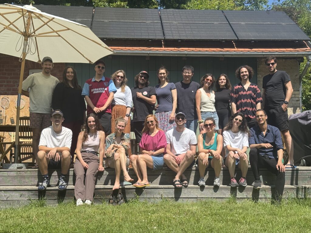
A few weeks later we gathered for our yearly retreat at Forsthaus Tornow. Alongside discussions about ongoing and future projects, there was plenty of time to enjoy the surroundings — swimming in the lake, sitting by the fire, and long walks through the forest.
We’re already looking forward to repeating the running and the resting in 2026.