

Our recent study “Fractal analysis of brain shape formation predicts age and genetic similarity in human newborns” has been published in Nature Neuroscience.
Background:
In this work, we studied how the human brain develops over the first few weeks after birth. While it is well known that the brains of newborn babies increase rapidly in size, here we studied how the brain develops its shape (see Fig. 1 below).
To do this, we analyzed structural MRI data of nearly 800 human newborns from the developing Human Connectome Project (dHCP) and measured their brain shape with fractal dimensionality (FD) – a geometric measure of structural complexity.
Key results:
- Brain shape reflects infant maturity better than brain size: FD was consistently better at capturing infant age compared to brain size (measured by volume). Not least, we were able to predict the age of the babies from their brain shapes with a mean error of ~4 days, significantly outperforming age prediction from brain size.
- Brain shape detects traces of premature birth that are not captured by brain size: Prematurely born infants showed persistent alterations of FD but not volume compared to term-born babies, even when they were scanned at the same age.
- Similarity in brain shape reflects similarity in genetic information: The brains of any two babies were more similar in shape if they shared more genetic information. Specifically, brain shapes were more similar in infants of the same sex, in biologically related compared to unrelated infants, and in identical twins (that share ~100% of their genes) compared to fraternal twins (that share ~50% of their genes).
- Brain shape analysis with FD systematically outperforms previous measures: Age and genetic information were captured systematically better by FD – not only compared to volume but also to other common brain measures, including cortical thickness, curvature, gyrification, sulcation, surface area, and the T1-weighted/T2-weighted ratio.
Interpretation:
The formation of brain shape represents a fundamental maturational process in human brain development – beyond growth in size. Brain shape analysis with FD captures biologically relevant information better than earlier neuroimaging measures, offering a powerful new framework to study both normative brain development and disease-related changes of brain morphology.
Like to know more?
You can find the full paper here (open access).
The study was highlighted in a paper spotlight by the Charité press department (in English and in German) and was voted paper of the month at Charité Neurology.
If you’re interested, do reach out to us – we are always happy to discuss!
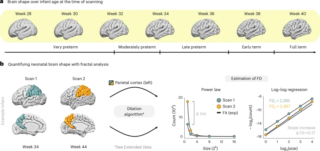
Fig. 1: Quantifying brain shape in human newborns. a, Differences in brain shape over infant age at the time of scanning, illustrated for cortical gray matter of the left hemisphere. Surface renderings correspond to the age-specific group averages of the developing Human Connectome Project (dHCP). Maturity levels follow the criteria by the World Health Organization (WHO) and the American College of Obstetricians & Gynecologists (ACOG). b, Quantifying neonatal brain shape with fractal dimensionality (FD). The FD estimate is calculated from a dilation procedure of the voxel-indexed segmentation mask, which measures the scaling properties of the structure through iterative convolution with varying spatial kernels (see Extended Data Fig. 1 for an illustration). Scaling behavior is assessed by the power law relationship between kernel size and the count of scaled measurement units after convolution. The slope of this relationship in log−log space then yields the structure’s FD estimate. This estimation is illustrated for the left parietal cortex of an exemplary infant born at 32.6 weeks and scanned at 34 weeks and 44 weeks post-menstrual age. Over this 10-week interval, the morphological change of the region (left) is reflected by an increase in the structural complexity estimate (right). exp, exponential; Vol, volume.